Model of osteoporosis - 3B Germany
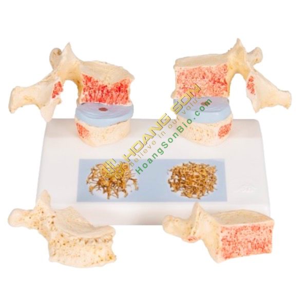
- Impressive didactic model to compare normal and osteoporotic thoracic vertebrae.
- Ideal for medical research and patient consultation. The 11th and 12th thoracic vertebrae are shown.
- Reconstruction of sequential osteoporotic thoracic vertebrae with narrower intervertebral disc located to the left of the bracket.
- The upper vertebrae are divided in the middle. The magnetically attached vertebral halves can be easily removed to show cutting surfaces.
- This allows a clear visualization of the fractured upper part of the vertebral body due to sintering, i.e. the breakdown of the bone substance during this process and the resulting osteoporosis.
- Degenerative changes in bone, expressed in the form of osteoclasts, are also recognizable.
- For comparison, copies of two healthy vertebrae corresponding to the intervertebral disc are provided on the right side. Half of the upper vertebral body is magnetically attached and can be removed.
- The detailed illustration on the base depicts two 3D micro-CT images obtained from bone biopsies.
- These illustrate the microarchitecture of osteoporotic bone, which has lower bone density than healthy bone.
- Specifications:
+ Weight: 0.834 kg
+ Dimensions: 26 x 19 x 14.5 cm
- Brand: 3B Scientific